Veterinary surgeons only
Neurology & Neurosurgery
Neurology is a branch of veterinary medicine dealing with disorders of the nervous system. Our veterinary referral neurology team is trained to investigate, diagnose and treat medical and surgical disorders involving the central and peripheral nervous systems. Our team is also involved in clinical research and national and international teaching activities.
Our neurology team consists of four European and RCVS Veterinary Specialists in Neurology and a neurology intern. (for more information about European Specialists in Veterinary Neurology please click here)
With onsite MRI, CT and an electrodiagnostic system we are well equipped to manage neurology and neurosurgical cases. Our neurology team is supported by other speciality services, interns and nursing team and this allows pets to receive a state-of-the-art round-the-clock care throughout their hospital stay.


Our on-site MRI and CT scanning is performed by our nurse technicians who are trained in acquiring images of the highest quality. One of our imaging technicians has been awarded the status of Veterinary Magnetic Resonance Imager (RVMRI) with the American Association of Veterinary Radiologists (AAVR).
The neurosurgical unit is supported by first rate nursing care and 24 hour patient supervision by veterinary interns. The service is also supported by veterinary physiotherapists. We are one of the few veterinary hospitals using a neurosurgical microscope for spinal and brain surgery in small animals. The surgical microscope features brilliant illumination and customisable options to offer the quality and reliability needed to perform outstanding neurosurgery in small animals.


The video shows the surgical stabilisation in a dog with incomplete ossification of the atlas.
The video shows the surgical stabilisation in a dog with congenital atlanto-axial subluxation.
Below you can watch videos showing surgeries performed with our neurosurgical microscope. WARNING: these videos contain intraoperative images that may offend the viewers.
This video shows a spinal surgery for the removal of a subarachnoid diverticulum in a young pug performed with our neurosurgical microscope.
This video shows a spinal surgery for the removal of a lateralised extruded intervertebral disc at the cervical spine in a dog performed with our neurosurgical microscope.
This video shows a de-bulking surgery for the removal of a large neoplasm (transitional meningioma) located at the lumbar spinal cord in a Jack Russell Terrier performed with our neurosurgical microscope.
This video shows the surgical biopsy of the common peroneal nerve in a dog.
Neurological disorders that we commonly encounter in our small animal patients include:
- Epilepsy
- Meningitis and meningoencephalitis of unknown origin
- Spine and spinal cord malformations
- Spinal trauma (vertebral fracture and luxations/subluxations)
- Brain tumours
- Hydrocephalus
- Intervertebral disc disease
- Infectious diseases of the nervous system
- Neuromuscular disorders.
Would all the diagnostic investigations take place on the same day as the consultation?
Commonly, we need to carry out a step-by-step diagnostic investigation approach which could take a few days. Where possible, we may carry out all the diagnostic investigations on the same day. Please remember that we are a busy multidisciplinary hospital seeing many emergencies. Emergency cases must take priority, which can impact the schedule for other patients – we know that you would understand if your own pet was in this situation.
What are the usual clinical steps that the neurologists will carry out to find out what is wrong with my pet and to find a solution?
During the consultation, the neurologist will firstly ask you a few questions to better understand whether your pet is affected by a neurological condition and to broadly define which neurological disease your pet is likely to suffer from. The neurologist will then carry out a complete neurological examination and will discuss with you the suggested diagnostic process which may include blood tests, diagnostic imaging (MRI and/or CT scanning, ultrasonography), cerebrospinal fluid analysis, electrodiagnostic investigations. After the diagnosis has been formulated, the neurologist will have a conversation with you about therapy options and prognoses.
After the diagnostic procedures, would I be able to take my pet home?
Pets are sometimes discharged on the same day as the diagnostic procedures. However, if it is in your pet’s best interest to remain hospitalised for observation or recovery, this is what we will advise. Please be reassured that during the hospital stay your pet will be looked after our nursing, interns and specialists team.
If my pet is hospitalised, will I be contacted to let me know how he/she is doing?
Yes, we will contact you on a daily basis. Our standard is that you will receive an update every weekday morning and evening, and once daily during weekends while your pet remains hospitalised. This will be from a member of the neurology team – either a clinician or nurse.
Will my pet be discharged by the clinician responsible for the case?
Usually, the clinician who has performed the work-up on your pet will discharge him/her. However, it may be that your pet is discharged by another clinician or one of our nurses. In the latter case, a clinician will ring you to discuss the diagnosis and therapy options for your pet.
Would I be notified about the results of the diagnostic investigations on the same day of the investigations?
The clinician may be able to discuss some of the findings of the diagnostic investigations with you on the same day. However, depending on the complexity of the case, we would usually need a few days or weeks (often no more than 2-3 weeks) to acquire all of the results and to formulate a presumptive or definitive diagnosis, to plan therapy and to notify your referring veterinary surgeon.
How do I get my pet referred to NWVS?
You can request referral to our neurology team by speaking to your vet.
Is MRI an invasive imaging technology?
MRI is a non-invasive imaging technology that produces detailed anatomical images without the use of ionising radiation.
How long does an MRI examination take?
Full examination of the brain in small animals normally takes approximately 45-60 minutes. The time to carry out a full examination of one portion of the spine may vary, but it usually takes around 60 minutes.
Is CT an invasive imaging technology?
CT scans are generally safe. However, your pet will exposed to X-ray radiation. CT scanners are designed to make sure that your pet is not exposed to unnecessarily high levels of X-ray radiation.
How long does a CT examination take?
Commonly, CT examination takes only a few minutes to acquire the images.
Does my pet need an anaesthetic for these procedures?
It is really important that the patient is still to acquire images for both MRI and CT. Anaesthesia is therefore essential for MRI. Sedation may be appropriate for CT but will always be at the discretion of our specialist anaesthetists.
Testimonials
What our clients say:
 Vincent Ward2024-04-02An exemplary practice in all respects. The veterinary surgeons, nurses and support staff continue to take such wonderful care of my Labrador, Albie. He could not be in safer hands. I have no hesitation whatsoever in recommending this practice to all those seeking specialist medical treatment for their beloved pets.John Taylor2024-03-27Caring staff, saved my dog's lifeLisa Braithwaite2024-03-25Absolutely incredible team, everyone was extremely friendly and the vets were amazing. So knowledgable and communicated everything to us about our dog. The treatment our dog received was amazing and when we picked him up we had a full care plan going forward. I would 100% recommend if your pet needs specialist treatment!Vicki Lester2024-03-23Absolutely fantastic service. From the receptionist to the vets and nurses, they were all brilliant. Made a very stressful time easier for us. Very grateful for their compassion and support in making the right decisions for our little BellaPam Adshead2024-03-17Cannot praise them highly enough. They saved Harry’s life, everybody from reception through to Serena were incredibly kind and compassionate. Thank you doesn’t cover itRosie J2024-03-08Bruno was treated amazingly, like he was one of your family, thanks!Ciara2024-03-04Wow what an amazing practice! Such a welcoming environment with very friendly receptionists. We saw Francesca today whose knowledge is phenomenal, and she really took the time to talk in depth about our concerns and to get a very in depth history. She even went out of her way to get a neurologist to look at our dog to give us peace of mind. Blood tests done same day no issues. Highly recommend. Thank you Northwest!Bob Neale2024-02-18From arrival at the hospital under very traumatic circumstances we were supported and reassured that we were in the best place possible for our pet dog to recover from her injury. Everyone we met was understanding, empathetic, very reassuring and this was a great help. "Thank you" is not enough to express our level of appreciation.


Arranging a Referral or Case Advice
If you are a veterinary professional and would like to make a referral, or require pre-referral advice about a patient, please use the online forms.
Neurology Service Facilities
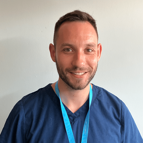
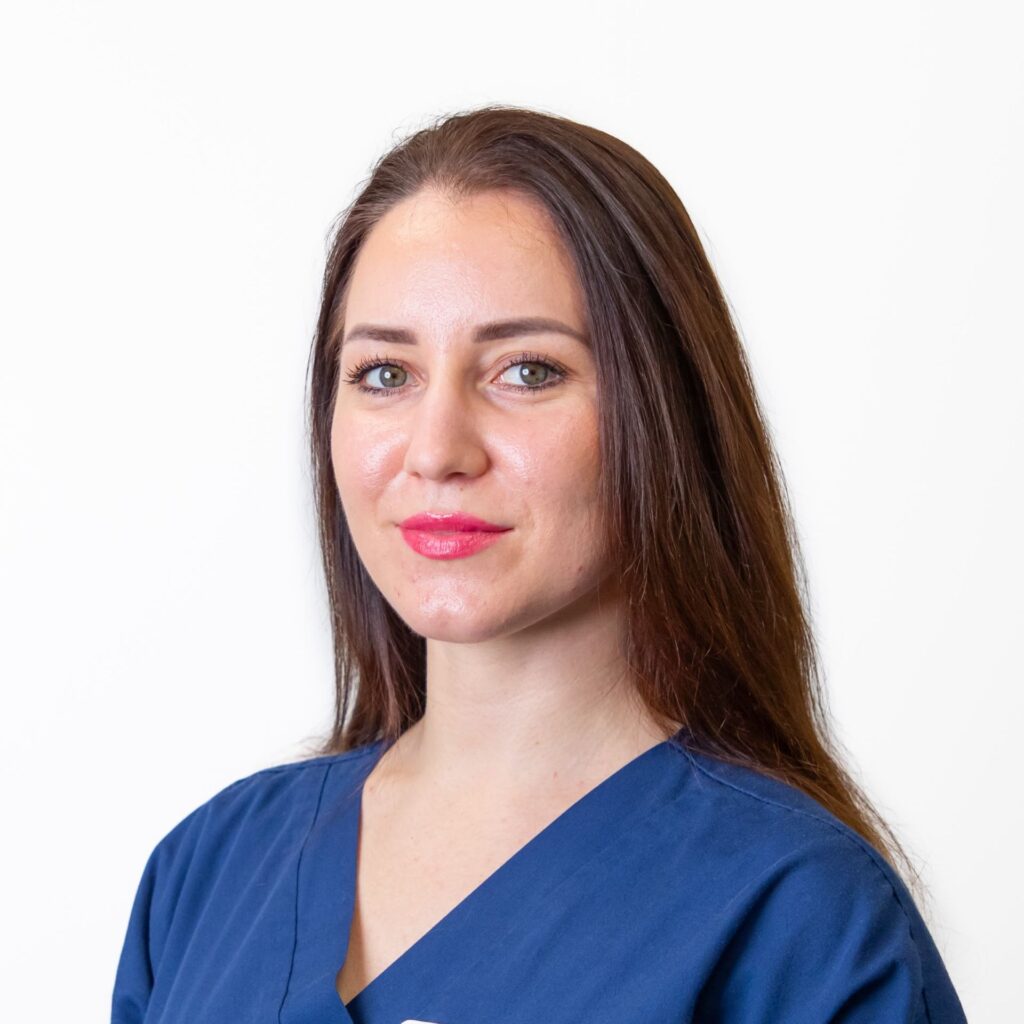
Meet the Team
Our specialist team working in this department
Selected published literature
-
Lorenzo Golini, Luca Motta, Massimo Mariscoli (2018). The operative microscope in modern neurosurgery. In Practice. 50(8), pp 20-21
-
Saulnier Troff F.G., Motta L., De Busscher V. (2017). Commentary: Complete Cranial Iliac Osteotomy to Approach the Lumbosacral Foramen. Frontiers in Veterinary Science 4:106
-
Mandara MT, Motta L, Calo’ P. (2016) Distribution of feline lymphoma in the central and peripheral nervous systems. The Veterinary Journal 216, 109–116
-
Motta L (2016). Does medical therapy influence the size of the syrinx in dogs with Chiari-like malformation/syringohydromyelia complex? Journal of Small Animal Practice 57, 278.
-
Motta L., de Lahunta A. (2015) Canine hemifacial spasm: a misnomer? Journal of Small Animal Practice, 56, 480
-
Ferreira, A, Sottiaux, J, Mandara, M. T., Motta, L. (2015) Ascending haemorrhagic myelomalacia associated with systemic hypertension in a hyperthyroid cat. Journal of Feline Medicine and Surgery. Open Reports 1–6
-
F.G. Saulnier-Troff, V. De Busscher, L. Motta. Improved exposure of the lumbosacral foramen using iliac osteotomy: preliminary results in 6 dogs with severe foraminal stenosis. Proceedings: 17th ESVOT Congress 2014, Venice (Italy), 2nd – 4th October, pp. 363-364
-
Motta, L. and Dutton, E. (2013). Suspected exercise-induced seizures in a young dog. Journal of Small Animal Practice, 54, 213-218
-
Gredal, H., Toft, N., Westrup, U., Motta, L., Gideon, P., Arlien-Søborg, P., Skerritt, G.C. & Berendt, M. (2013) Survival and clinical outcome of dogs with ischaemic stroke. The Veterinary Journal Jun;196(3):408-13
-
Motta, L. and Skerritt, G. C. (2012). Syringosubarachnoid shunt as a treatment for syringohydromyelia in dogs. Journal of Small Animal Practice, 53, 205-212
-
Motta, L., Skerritt, G. C., Denk, D., Leeming, G., and Saulnier, F. (2012). Dermoid sinus type IV associated with spina bifida in a young Victorian Bulldog. Veterinary Record, 170, 127-129
-
Motta, L., Mandara, M. T., and Skerritt, G. C. (2012). Canine and feline intracranial meningiomas: an updated review. The Veterinary Journal, 192, 153-165
-
Motta, L., Michal Altay, U., and Skerritt, G. C. (2011), Bell’s palsy with concomitant idiopathic cranial nerve polyneuropathy in 7 dogs. Journal of Small Animal Practice, 7: 397
-
14. Motta, L., Michal Altay, U., Kelly, D., and Skerritt, G. C. (2011). Intracranial Germ Cell tumour in an Airedale Terrier dog. European Journal of Companion Animal Practice. 21(2), 179-183.
-
Motta, L., Michal Altay, U., Carmichael, N., and Skerritt, G. C. (2011). Spinal haemorrhage associated with factor VII deficiency in a young Beagle. European Journal of Companion Animal Practice 21(1), 68-71
-
Motta, L., Michal Altay, U., Kelly, D. and Skerritt, G. C. (2011), Non-enhancing confirmed oligodendroglioma in three dogs. Journal of Small Animal Practice, 52: 227
28. Michal Altay, U., Motta, L., Woolley, J., and Skerritt, G. C. CLINICAL SIGNS IN ASSOCIATION WITH RATHKE’S POUCH CYST IN 11 DOGS. Abstract. Journal of Veterinary Internal Medicine, Feb 2010, 245
29. Vicens Zanoguera L, Pauciulo C, Corlazzoli D, Cauduro A, Motta L. Does surgical timing affect the rapidity of recovery in deep pain-entire non-ambulatory dogs with thoracolumbar intervertebral disk extrusion? J Small Anim Pract. 2022 Oct 31. doi: 10.1111/jsap.13570.
30. Compagnone K, A Upchurch D, Pompermaier E, Motta L. Thoracolumbar Intervertebral Disk Extrusion in Dogs: Do Onset of Clinical Signs, Time of Surgery, and Neurological Grade Matter? Vet Comp Orthop Traumatol. 2023 Jul 5. doi: 10.1055/s-0043-1770355.