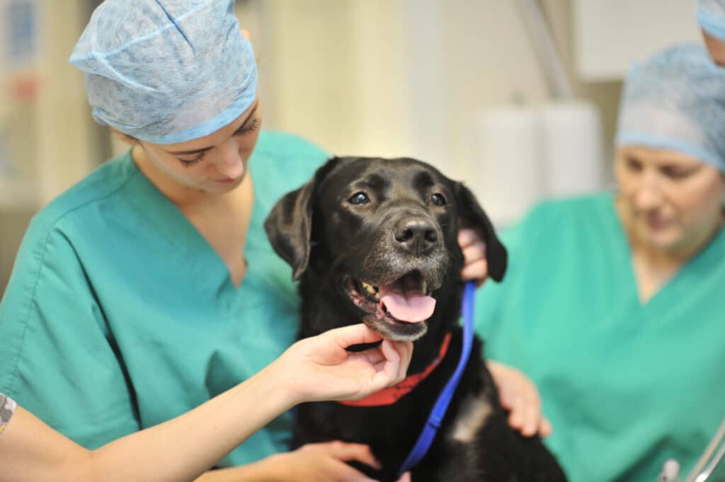
Beautiful Bella recovers from mast cell tumour removal
19 August 2020 / General
Bella is a beautiful 5-year-old black Labrador who belongs to Jenny Pearson – Support Services Manager at Northwest Veterinary Specialists. Jenny had recently noticed a mass on Bella’s left flank which seemed to have gone up and down in size. She was otherwise very well in herself.
On clinical examination with us in Runcorn, there was a 45mm subcutaneous mass located on her left cranial thoracic wall just caudal to the axilla. The regional lymph nodes were not palpable. No other masses were found. No abnormalities were found upon thoracic auscultation and she was comfortable on abdominal palpation.
A fine-needle aspirate was taken from the mass and stained with Diff-Quik. We examined the slide under the microscope in-house. This showed a population of small-to-medium-sized round cells with abundant, small, uniform cytoplasmic granules which stained purplish red (fig 1). This appearance is characteristic for a mast cell tumour.

Staging was performed to assess for any evidence of metastasis or co-morbidities prior to surgery. Routine blood work was unremarkable. Thoracic radiographs showed no pre-sternal lymph node enlargement. On abdominal ultrasound, there was no detectable lymphadenopathy and the liver and spleen were both homogenous and displayed respective normal sonographic features. Given that the regional lymph nodes (left prescapular and axillary) were not palpable, these were more accurately assessed with ultrasound, for palpably normal lymph nodes can still be metastatic. In Bella’s case, the lymph nodes were considered to be normal in size (fig 2). Ultrasound guided fine-needle aspirates were taken from the lymph nodes, spleen and liver and showed no evidence of metastasis. Chlorphenamine (H1 blocker) was administered prior to aspiration and she was discharged with further chlorphenamine and omeprazole (proton pump inhibitor) to reduce the potential for systemic effects associated with mast cell degranulation.

Bella was then transferred to our soft tissue service for excision of the mast cell tumour (fig 3 and 4). The tumour was excised with 20mm gross lateral margins and 2 fascial planes deep. The incision was performed through the lower edge of latissimus dorsi, cutaneous trunci and dorsal edge of pectoralis, and fat on the caudal aspect of triceps. Reconstruction of the surgical site was planned and performed with a sub-dermal plexus rotational flap. This particular flap was created from the dorsal thoracodorsal domain with an incision curving dorso-cranially off the top of the incision. At closure there was good range of motion from the elbow.


The subsequent histopathology report confirmed the diagnosis of a subcutaneous mast cell tumour which had been completely excised with 12mm deep and 15mm lateral margins. It was classified as low grade (Kiupel) with a mitotic index of 1/10hpf. The Patnaik grading system which is used for cutaneous mast cell tumours is not appropriate for subcutaneous tumours.
Bella made an excellent full recovery from surgery with no postoperative issues.
We advised Jenny that in the case of subcutaneous mast cell tumours, these typically have a less aggressive behaviour with a more favourable prognosis and extended survival times. In one study of 306 dogs with subcutaneous mast cell tumours, the metastatic rate was only 4% and only 8% experienced local recurrence. The estimated 2 and 5 year survival probabilities were 92% and 86% respectively. So the majority of patients can have a good outcome following surgery. There is a subset of these tumours which can have a more aggressive behaviour and this can be associated with a mitotic index of greater than 4 and a more infiltrative growth pattern. In Bella’s case, given the complete margins and low mitotic index, we would expect the surgery to be curative without the need for adjuvant treatment, although active monitoring for local recurrence would still be advised. Bella may be predisposed to developing more mast cell tumours in the future and so Jenny will continue to monitor for any new masses.


